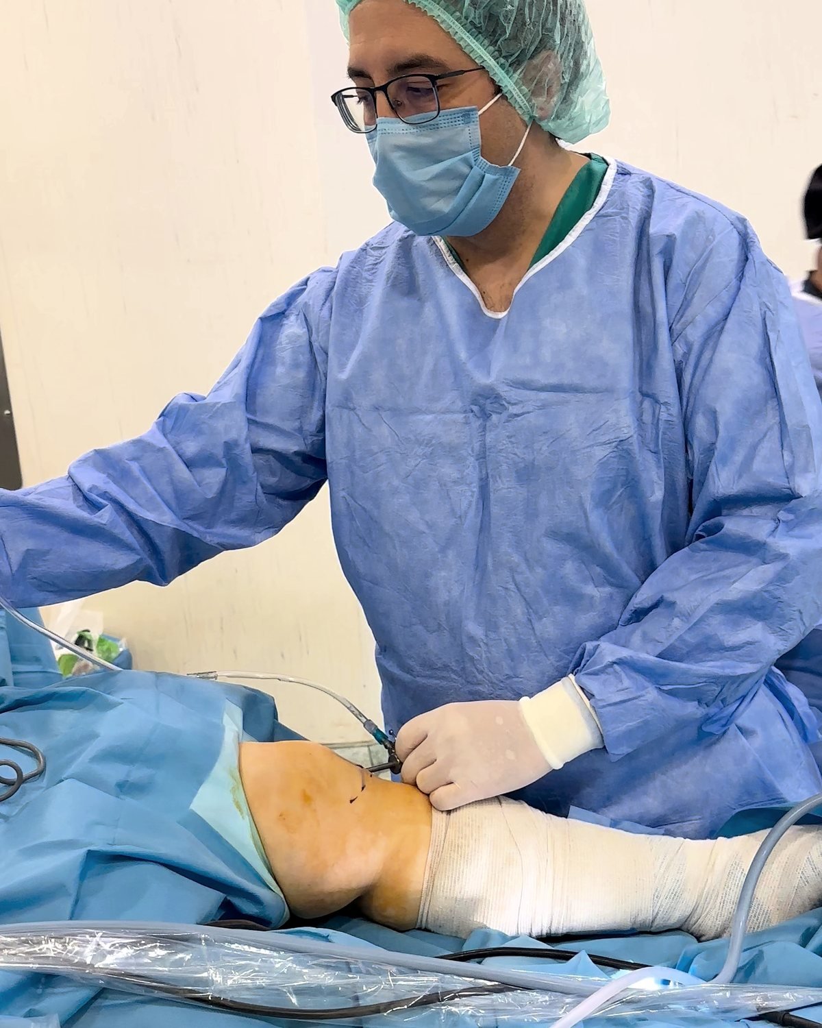
What is an arthroscopy?
Inside a joint, the ends of the bones are covered with a smooth, shiny layer of cartilage. This layer lets the joint move easily, without friction. There are also soft tissues: in the knee, the menisci (cushions of cartilage) and the cruciate ligaments; in the shoulder, the rotator cuff (the tendons of the muscles that rotate the shoulder) and the labrum (a rim of cartilage around the socket).
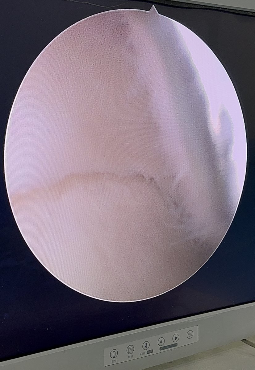
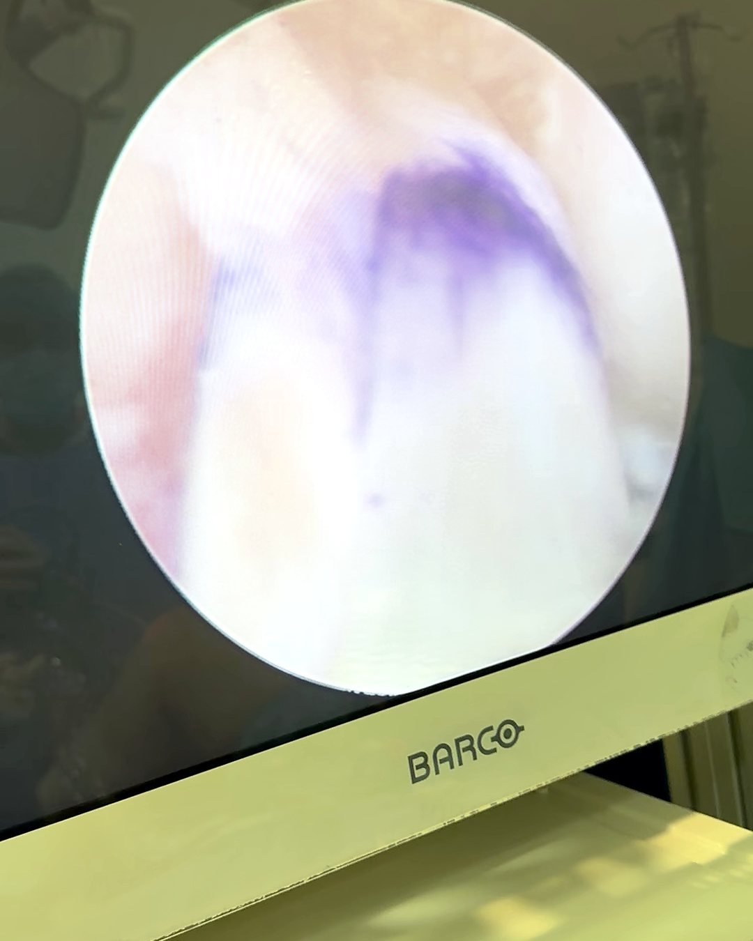
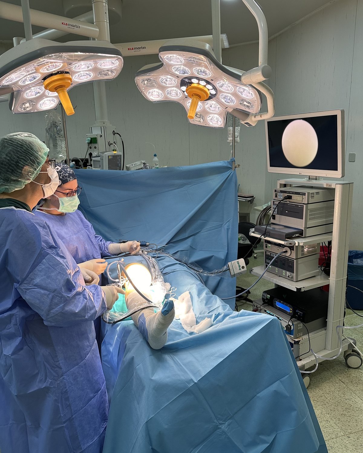
In an arthroscopy, we make two to four small cuts of about 1 cm around the joint. A camera goes in through one cut and fine instruments through the others. The inside of the joint is shown enlarged on a screen, and the necessary repair is carried out.
Joint cartilage has a very limited ability to repair itself. Limited cartilage damage can follow a sports injury, a sprain or a problem with the blood supply to the bone under the cartilage. With an arthroscopy, this damage can be assessed and treated before it progresses to osteoarthritis.
When is an arthroscopy done?
The decision is based on the examination and, in most cases, an MRI scan. Not every tear needs an operation. We consider the type of your symptoms, your age and your activity level together.
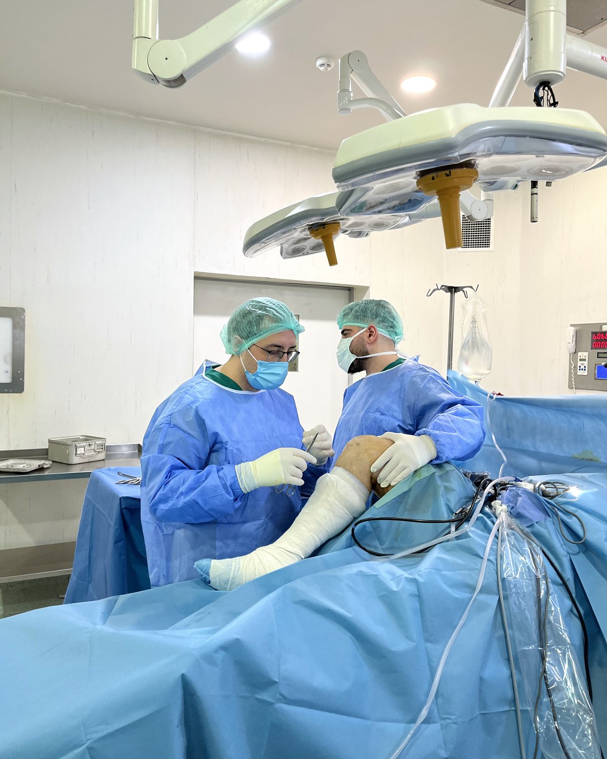
Knee arthroscopy
- Meniscus tear: tears that can be stitched are repaired; damaged parts that cannot be stitched are trimmed in a limited way
- ACL and PCL (anterior and posterior cruciate ligament) tears: a new ligament is made from your own tendon
- Cartilage damage: cartilage repair, microfracture, cartilage transfer (mosaicplasty), and removal of loose fragments floating in the joint
- Kneecap instability, and problems that make the knee catch or lock
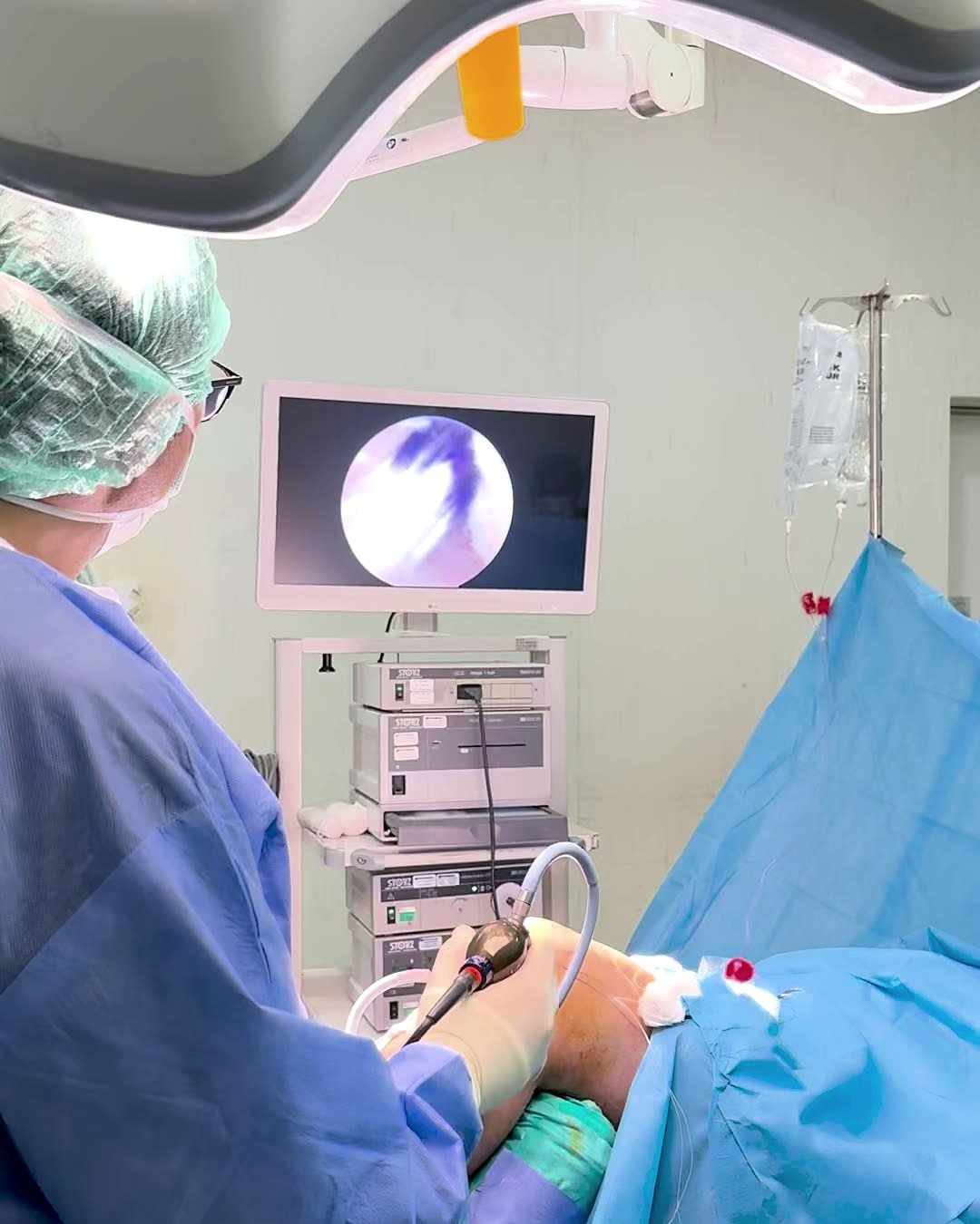
Shoulder arthroscopy
- Rotator cuff tear repair
- Labrum repair for repeated shoulder dislocations
- Shoulder impingement and calcium deposits in the shoulder
- Releasing the joint capsule in frozen shoulder that has not responded to medicines, injections and physiotherapy
Ankle arthroscopy
- Impingement (pinching) at the front or back of the ankle
- Cartilage damage of the ankle bone (talus): repair, microfracture and removal of loose fragments
- Inflamed tissue left behind by old sprains
How are cartilage problems treated?
Treatment of cartilage damage depends on its size, depth and position, and on your age and activity level. Osteoarthritis that has spread across the joint is different from cartilage damage limited to one area. In osteoarthritis the aim is to manage pain. In limited damage the aim is to repair the cartilage surface.
Recovery after cartilage repair takes longer than after meniscus surgery. Weight-bearing on the leg is limited for a few weeks so that the repaired tissue can mature, and rehabilitation moves forward in stages with the physiotherapist. We do not recommend an arthroscopy only to "clean out" a knee with widespread osteoarthritis.
- Cartilage trimming: loose flaps of cartilage that cause catching are removed and the edges of the damage are smoothed.
- Microfracture (bone marrow stimulation): for small areas of damage, tiny holes are made in the bone under the cartilage. The area fills with repair tissue formed by cells from the bone marrow.
- Cartilage transfer (mosaicplasty): small plugs of cartilage and bone are taken from a part of the knee that carries little weight and placed in the damaged area. It is used for medium-sized damage and sometimes needs a small open cut.
- Fixing a detached fragment: especially in young people and athletes, a fragment of cartilage and bone that has come loose can be put back and fixed if it is suitable.
- Membrane-supported repair: for larger damage, biological membranes that support the repair tissue and cell-based methods may be considered.
Advantages and limits of keyhole surgery
Because the cuts are small, pain, bleeding and wound problems after surgery are usually less than after open surgery. The scars are small and the hospital stay is short. The camera shows the inside of the joint enlarged, so small damage that is not clear on MRI can also be assessed during the operation.
However, not every problem can be solved with keyhole surgery. For large bone loss, some fractures and severe osteoarthritis, open surgery or a joint replacement may be more suitable. Before the operation, we discuss openly which method suits you.
Getting ready and the day of surgery
You have routine tests before surgery, and we review the medicines you take. For suitable patients, physiotherapy starts before the operation to reduce swelling and keep muscle strength (prehab).
Knee and ankle arthroscopy is usually done under spinal anaesthetic (numbing from the waist down). Shoulder arthroscopy is usually done under general anaesthetic, with an ultrasound-guided nerve block for the shoulder added. Depending on the procedure, the operation usually takes between 30 minutes and 2 hours. Most patients go home on the same day or after one night in hospital.
What is recovery like?
Dr Gürsel and the physiotherapist see you together at your follow-up appointments. The small cuts make it easier to start moving early, but the repair inside the joint still needs time to heal.
After simple meniscus trimming, most people walk within a few days and usually return to daily activities within 2–6 weeks. After a meniscus repair, ligament surgery or a shoulder tendon repair, crutches or an arm sling are used for a few weeks, and rehabilitation takes months.
Possible risks and side effects
Although it is done through small cuts, an arthroscopy is still an operation. Complications are rare but can include infection, blood or fluid building up in the joint, a blood clot in the leg veins, nerve injury and joint stiffness.
Contact us without delay if you have a fever, discharge from a wound, increasing swelling and redness of the joint, or sudden swelling of the calf.
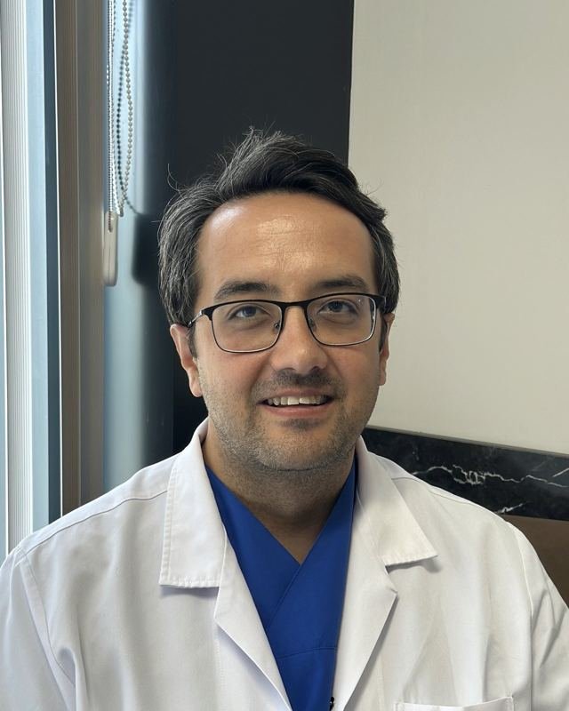
Reviewed by Dr Enis Gürsel, Orthopaedic and Trauma Surgeon · Last reviewed: 5 November 2026