Are a heel spur and plantar fasciitis the same thing?
The pain that people call a heel spur usually comes from plantar fasciitis. This is inflammation and wear where the thick band of tissue on the sole of the foot (plantar fascia) attaches to the heel. The plantar fascia works like a spring that supports the arch of the foot, and it is strained by repeated load.
A heel spur is a small bony point that grows from the heel bone and shows on an X-ray. Many people have a heel spur without any pain, and many people with pain have no spur at all. So we aim the treatment at the painful tissue, not at the spur itself.
What are the symptoms, and who gets it?
Typically the heel hurts in the first steps of the morning and when you stand up after sitting for a long time. The pain eases after a few steps and can come back at the end of the day. Being overweight, standing for long hours at work, running on hard surfaces, flat feet, tight calf muscles and unsupportive shoes increase the risk.
Heel pain does not always come from plantar fasciitis. Achilles tendon problems, a stress fracture of the heel bone, a trapped nerve in the tarsal tunnel (on the inside of the ankle) or rheumatic diseases can cause similar symptoms. That is why we use examination and ultrasound to confirm the source of the pain before treatment.
First step: shoes, insoles and stretching
With these measures, many people get relief over weeks to months. What matters is doing them regularly. Our physiotherapist, Gökçenaz Arpaçay, shows you how to do the exercises correctly.
- Wear shoes with a soft heel and arch support, and avoid walking barefoot at home too
- Use a silicone heel cup or an insole that suits your foot
- Do calf and sole stretches, including before you get out of bed in the morning
- If needed, a night splint, weight control and short-term painkillers
What if this is not enough?
If the pain continues despite these measures, we can give an ultrasound-guided injection of local anaesthetic and steroid (cortisone) around the plantar fascia. We use steroid injections only a limited number of times, because they carry a risk of thinning the heel fat pad and tearing the fascia. In suitable patients, PRP (platelet-rich plasma, prepared from your own blood) is another option.
If an injection does not help, or the pain keeps coming back, we use radiofrequency treatment. With ultrasound we can see the small nerve branches that carry pain from the heel (branches of the medial calcaneal nerve and Baxter's nerve) and the painful attachment of the fascia, and we treat them with a needle. The aim is to reduce pain for a long period, so that exercise and daily walking become easier.
How do we keep the treatment comfortable?
The skin and fat pad of the heel are among the most sensitive areas of the body. Injections given directly into the heel are usually very painful, and many people avoid treatment for this reason.
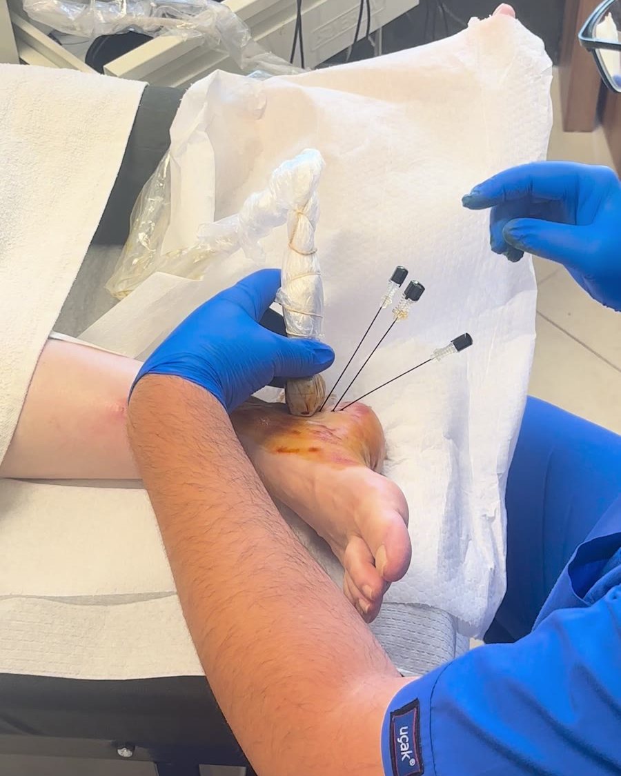
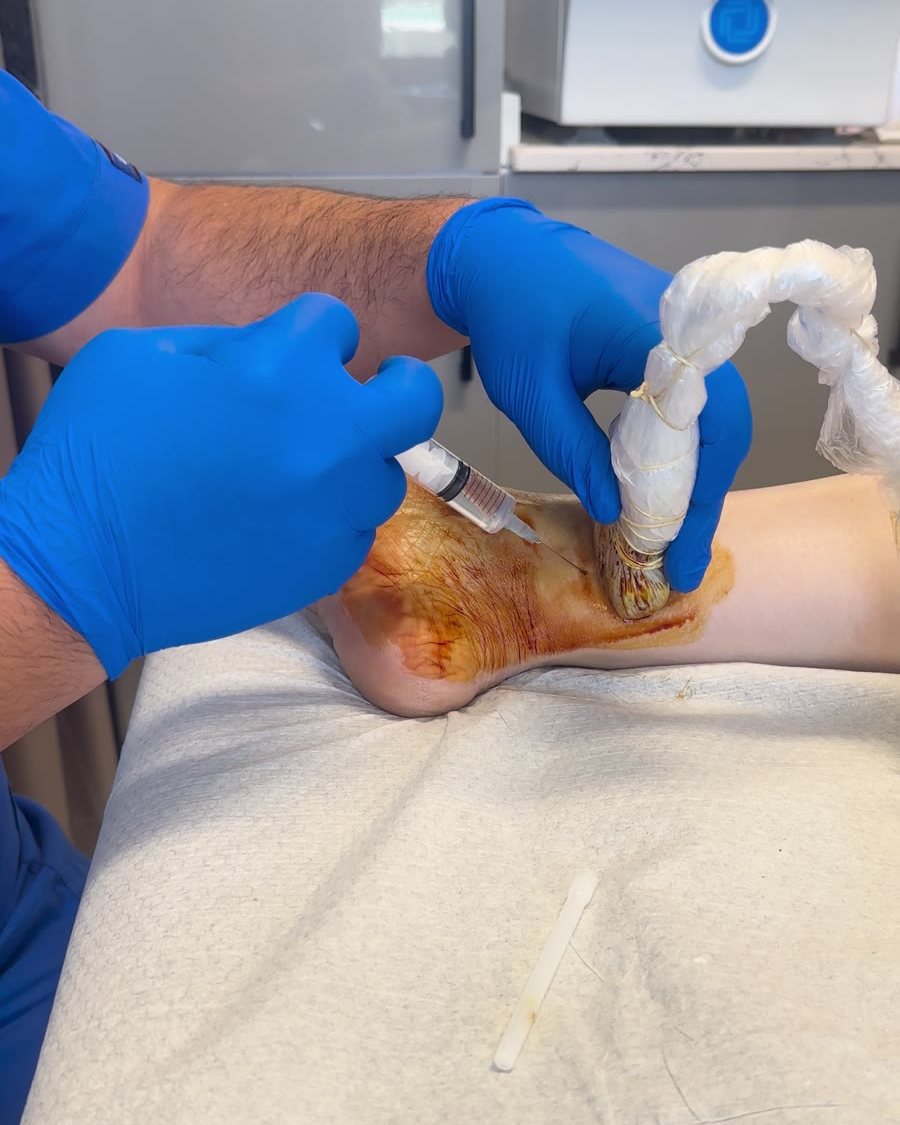
Before we start, we numb the nerves that carry feeling to the heel. We do this from the inside of the ankle with a fine needle, under ultrasound guidance (a nerve block, which is a local anaesthetic injection around a nerve). You feel this much less than an injection into the heel. Within a few minutes the heel becomes numb, and only then do we give the injection or the radiofrequency treatment.
In this way, the aim is that you do not feel pain during the treatment. The numbness in the heel can last for a few hours. During this time, walk carefully and do not drive.
Why use ultrasound?
Ultrasound shows the thickened fascia and the painful spot. It lets us guide the needle to the target tissue rather than into the heel fat pad. It also supports the diagnosis, because it helps us tell apart tendon problems, trapped nerves and bone problems that can also cause heel pain.
What happens on the day of treatment?
You do not need to fast beforehand. Please come in comfortable shoes that support your heel. If you take blood-thinning medicines, tell us in advance.
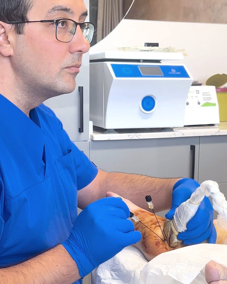
You lie on your front or on your side. First we numb the nerves to the heel from the inside of the ankle, using ultrasound. When the heel is numb, we give the injection or the radiofrequency treatment. The procedure usually takes 20–30 minutes.
Afterwards you can walk home. Until the numbness wears off, walk carefully and do not drive. For a few days, avoid long walks and running.
Possible risks and side effects
You may have a temporary increase in pain for 24–48 hours, bruising, and a change in feeling on the inner side of the heel. Infection, bleeding and nerve damage are rare risks. A steroid injection carries a risk of thinning the heel fat pad and tearing the fascia.
After treatment: exercise and follow-up
Radiofrequency treatment or an injection does not replace stretching exercises. At your follow-up appointment we check your pain and how you walk, and continue the exercise and insole plan. Very rarely, if the pain does not respond to any of these treatments, surgery may be discussed.

Reviewed by Dr Enis Gürsel, Orthopaedic and Trauma Surgeon · Last reviewed: 5 November 2026